

028 38 352640

Contact Us
Greenmount Veterinary Clinic
72 Gilford Road,
Portadown, Co Armagh,
BT63 5EG
Tel: 028
38 358525/352640

Ophthalmology Referrals & Cataract Surgery
Cataract Surgery
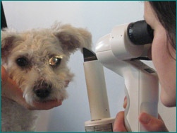
A referral Ophthalmology service is available at Greenmount vet clinic Portadown. With the aid of our advanced diagnostic and surgical eye facilities our Ophthalmologist Dr Isabel M. Buehler MRCVS is able to offer a wide range of additional services for pets with eye conditions, including onsite cataract surgery by phacoemulsification and intraocular lens implantation.
What is a cataract?
Cataract is a clouding of the lens in the eye. The lens is normally clear and transparent, and
its function is to focus light on the sensitive tissue at the back of the eye.
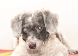
A cataract can affect just a small part of the lens, or it can affect the entire lens. If the
cataract involves the whole lens then vision will be affected. You may notice that the pupil,
which normally appears black, has changed colour and appears now bluish or white.
What is the treatment for cataracts?
Currently, the only proven technique to treat cataracts is surgery. There are unfortunately
no known preventative treatments for cataracts.
When is the best time to operate on a cataract?
In the past cataract surgery was usually delayed until the cataract had matured and the
patient had gone totally blind. We now know that surgery is considerably more successful,
if the cataract is removed before it matures.
It is therefore recommended to carry out cataract surgery as soon as the clouded lens starts
to interfere significantly with vision. This is especially important in young and diabetic
patients, where progression of cataracts can often be rapid and significant complications
are possible, if treatment is delayed.
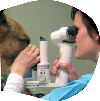
Why do we perform ultrasonography and blood tests before surgery?
As part of the general work-up we do blood tests to make sure your pet is free of any
problems that might interfere with the success of cataract surgery.
We also perform ocular ultrasonography. The ultrasound is used to image the inside of
the eye; in these cases we are checking to make sure that the eye is healthy enough for
surgery. In particular we are looking for advanced degenerations of the vitreous and/or
retinal detachments.
The surgery
Your pet will normally be admitted on the morning of the surgery. It is important to fast
your pet overnight, but water should not be withheld.
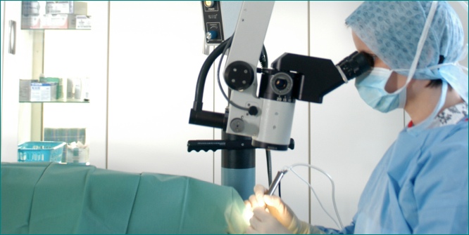
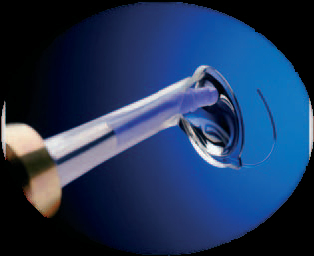
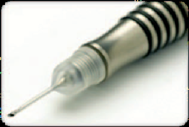
Cataract surgery is performed using the latest proven technique of phacoemulsification.
It is the same technique that is currently used in humans. Surgery is carried out under
general anesthesia with the aid of a phacoemulsification unit, an operating microscope
and tiny microsurgical instruments.
Two small incisions, the first 1mm, the second 2.8 mm long, are made into the eye and
part of the anterior capsule of the lens is taken out with a fine pair of forceps. The cataract
is then removed by phacoemulsification which is ultrasound energy used to break down
the cataract.
In most patients it is possible to insert a special artificial lens where the old lens was. Vision
will then be similar to the way it used to be before the surgery. The lenses used in our
patients are new generation foldable acrylic intraocular lens implants which are especially
made for pets.
Finally, the incisions are closed with fine hair like suture material.
What aftercare does my pet need?
The aftercare following cataract surgery will be intensive. Usually there are two types of
eye drops to be given several times daily. The number of applications though gradually
decreases over the following two to three months.
Most pets settle down quickly after the surgery, and there is generally little if any pain
associated with the procedure. A pain relief injection is given on the morning of surgery,
and your pet will also receive pain relief tablets for some time.
Your dog will need to be kept as quiet as possible for a few weeks after the surgery
(also no excessive barking or vigorous play allowed!!), although this can obviously be
challenging with some patients! No pulling on the lead will be allowed for several weeks
after surgery, as this puts up the pressure inside the eye and can encourage bleeding. It is
advisable to acquire a harness before the operation as this avoids pulling around the
neck. Also a plastic Elizabethan collar has to be worn for ten days after the operation.
There will be about five check-ups within the first few months after surgery. For some
patients long term treatment will be necessary, and they may require more re-examinations
than average.
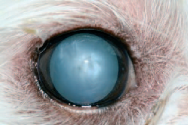
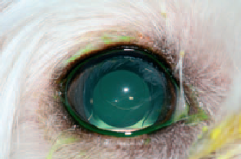
Pictures of a cataractous eye before and after cataract surgery
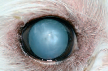
Before cataract surgery- opaque lens
After cataract surgery- clear vision
What possible risks and complications are there?
The success rate of cataract surgery in dogs is about 85 - 90%. Rarely, complications can
lead to the development of a painful and blind eye, and the removal of the eye may be
necessary.
Complications include inflammation (uveitis), glaucoma, retinal detachment, infection,
corneal edema, ulcers and wound breakdown. Some dogs have problems in their eyes
which cannot always be identified before surgery. This may mean the operation is not
successful or the vision may not be as good as envisaged.
The vast majority of patients are doing very well after their operation. If you decide to give
your pet the opportunity of a better vision, we will be delighted to give you as much help,
support and advice as possible.
If you have any queries or just would like to know more about cataract surgery, please do
not hesitate to contact us.
To make an appointment to see Ophthalmologist Dr Isabel M. Buehler MRCVS please contact the clinic on 028 38358525.
To make an appointment to see Ophthalmologist Dr Isabel M. Buehler MRCVS please contact the clinic on 028 38358525.

Website content owned and produced by Ewing Walker, Ewing may be contacted through Greenmount Veterinary Clinic or VetOrtho Referrals. Copyright © All rights reserved. Ewing Walker Greenmount Vets
| pet grooming |
| fleas & ticks |
| pet microchip |
| pet insurance |
| pet worming |
| pet neutering |
| vaccinations |
| dogs trust scheme |
| dog neuter for £15 |
| rabbits |
| cats |
| dogs |
| dental treatments |
| spinal xray myelogram |
| GSDA A stamp |
| elbow score |
| hip score |
| hip score guide for owners |
| our healthy pet offers |
| vetortho.co.uk |
| Fracture Surgery |
| Cruciate Surgery TTA TPLO |
| Hip Dysplasia |
| Spinal Surgery |
| Elbow Dysplasia |
| Cardiology services offered |
| Doppler heart scan |
| Blood pressure monitoring |
| Electrocardiogram ECG |
| Holter monitoring |
| Bronchoscopy & Endoscopy |
| ophthalmology referrals |
| cataract surgery |
| eye-vet |